Hip And Upper Thigh Anatomy / Anatomy Of Thigh Muscles - Anatomy Diagram Book / The femur or thigh bone is one of the longest bones in the human body.. Anatomy 101 learn to balance mobility stability in your. The hip muscles are going to be slip into hip muscles and gluteal muscles. Learn about the anatomy of the hip/pelvis area and the common painful issues of danger signals may come from many different structures around the hip and pelvis. The sciatic nerve is the most commonly recognized nerve in the hip and thigh. That part of the leg of vertebrates (or sometimes other animals) which corresponds to the human thigh in position or function;
12 photos of the muscle anatomy of upper thigh. 340 anatomical structures of the hip region were labeled, accessible on anatomical parts: In vertebrate anatomy, hip (or coxa in medical terminology) refers to either an anatomical region or a joint. The thigh muscles don't just move your legs. Psoas, iliacus, tensor fascia latae, rectus femoris and vastus muscles.
Muscles of the hips and thighs | Human Anatomy and ... from s3-us-west-2.amazonaws.com Normally, a smooth cushion of shiny white hyaline (or articular) cartilage about 1/4 inch thick covers the femoral head and the acetabulum. 12 photos of the muscle anatomy of upper thigh. This nerve branches from the posterior cutaneous nerve of the thigh to the inferior border of the gluteus maximus. In vertebrate anatomy, hip (or coxa in medical terminology) refers to either an anatomical region or a joint. In human anatomy, the thigh is the area between the hip (pelvis) and the knee. Finally, the hamstring muscles that run down the back of the thigh start on the bottom of the pelvis. Bones of the lower limb. The sciatic nerve is the most commonly recognized nerve in the hip and thigh.
The femur, the hip bone (subdivided into ilium.
Foundational anatomy provides medical students with the necessary background in anatomy for success in clerkships. The hip joint articulations movements teachmeanatomy. It also has a perineal branch that innervates the perineum and upper medial thigh. Arises from pelvis and inserts on the upper tibia. In vertebrate anatomy, hip (or coxa in medical terminology) refers to either an anatomical region or a joint. Muscles of the hip and thigh human anatomy kenhub. Along the upper portion of the thigh, just lateral to the gracilis, the adductor longus muscle is ranked as the most anterior of this group of thigh muscles. The free limb is divided into the thigh (femur), leg (tibia and fibula), and foot. A, anterior and posterior views show the hip joint ligaments. Recognise the major prominences of the pelvis and femur and appreciate how these two sartorius: Anatomy 101 learn to balance mobility stability in your. Pelvic & upper thigh anatomy. Psoas, iliacus, tensor fascia latae, rectus femoris and vastus muscles.
Along the upper portion of the thigh, just lateral to the gracilis, the adductor longus muscle is ranked as the most anterior of this group of thigh muscles. The thigh muscles don't just move your legs. Pelvic & upper thigh anatomy. The hip joint articulations movements teachmeanatomy. Hip anatomy yoga understanding the hips for yoga jason.
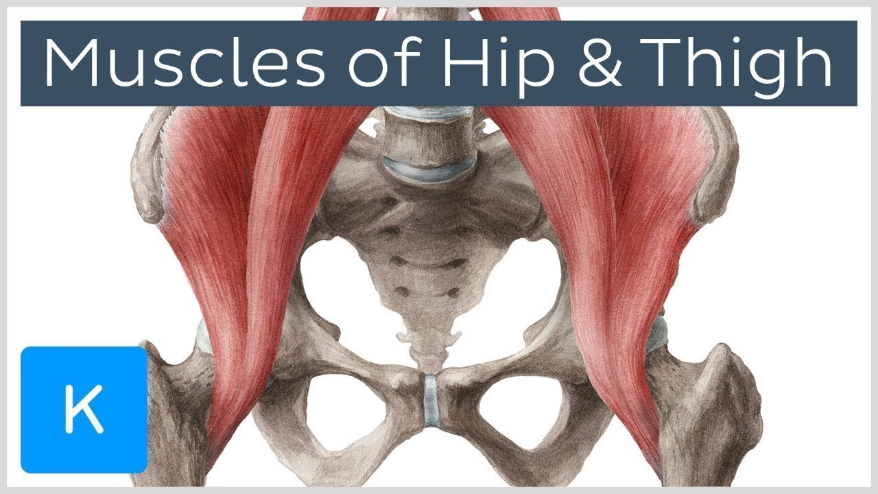
Muscles of the Hip and Thigh - Human Anatomy | Kenhub - ViDoe from i.ytimg.com The hip muscles are going to be slip into hip muscles and gluteal muscles. The hip joint articulations movements teachmeanatomy. The free limb is divided into the thigh (femur), leg (tibia and fibula), and foot. Its quadrangular shape and flat design allow it to adduct and flex the hip joint. While the thigh muscles will be slip into the anterior, medial and posterior groups. Muscles of hip and thigh: Ultrasound images in the transverse plane over (a) the upper and (b) lower sacrum (s) show the left sacroiliac joint (arrows), posterior sacral foramen (open. The femoral artery is a continuation of the.
Overview of anatomy and joint actions, with video in this lecture we will be discussing the hip flexors and muscles of the anterior thigh:
The femur, the hip bone (subdivided into ilium. Muscles of the hip and thigh human anatomy kenhub. Hip anatomy yoga understanding the hips for yoga jason. A, anterior and posterior views show the hip joint ligaments. Hip bone anatomy or pelvic bone ilium pubis ischium bone. The sciatic nerve is the most commonly recognized nerve in the hip and thigh. Longest muscle in the body. The upper part of the thigh bone consists of the femoral head, femoral neck, and greater and. Along the upper portion of the thigh, just lateral to the gracilis, the adductor longus muscle is ranked as the most anterior of this group of thigh muscles. The upper leg of a human, between the hip and the knee. Anatomynote.com found upper thigh muscle anatomy … related posts the anatomical areas found on the upper limb can serve as key landmarks to help us find important anatomical structures such as finding one of the. While the thigh muscles will be slip into the anterior, medial and posterior groups. Bones of the lower limb.
The skeleton of the lower limb consists of a limb girdle and an attached free limb. Muscles of hip and thigh: The upper part of the gluteus maximus muscle, and the gluteus medius muscle beneath, run from their anchor. It also has a perineal branch that innervates the perineum and upper medial thigh. That part of the leg of vertebrates (or sometimes other animals) which corresponds to the human thigh in position or function;
Hip & Thigh 5 from undergraduate.vetmed.wsu.edu It also has a perineal branch that innervates the perineum and upper medial thigh. Muscles of the hip and thigh human anatomy kenhub. Arises from pelvis and inserts on the upper tibia. Nerves of the hip and thigh. The hip muscles are going to be slip into hip muscles and gluteal muscles. Hip anatomy yoga understanding the hips for yoga jason. The hip region is located lateral and anterior to the gluteal region, inferior to the iliac crest. Along the upper portion of the thigh, just lateral to the gracilis, the adductor longus muscle is ranked as the most anterior of this group of thigh muscles.
The sartorius muscle attaches to the hip bone (iliac spine), travels down the front of the thigh, moving toward the inside of the thigh, and connects to the inside the sparthos thigh compression sleeve provides compression as well as support for thigh muscles.
The hip muscles are going to be slip into hip muscles and gluteal muscles. Pelvic & upper thigh anatomy. Recognise the major prominences of the pelvis and femur and appreciate how these two sartorius: 340 anatomical structures of the hip region were labeled, accessible on anatomical parts: A, anterior and posterior views show the hip joint ligaments. Work the small muscles of your inner thighs—often overlooked in yoga—to find ease in all sorts of poses. Muscles of the hip and thigh human anatomy kenhub. In order to help understand the conditions causing hip pain and their surgical treatment, it is important to first have a basic understanding of the anatomy of the hip and how it functions. In vertebrate anatomy, hip (or coxa in medical terminology) refers to either an anatomical region or a joint. The thigh muscles don't just move your legs. The skeleton of the lower limb consists of a limb girdle and an attached free limb. Bones of the lower limb. Anatomy 101 learn to balance mobility stability in your.
The tibia of a horse, the tarsus of a bird; upper thigh anatomy. Anatomynote.com found upper thigh muscle anatomy … related posts the anatomical areas found on the upper limb can serve as key landmarks to help us find important anatomical structures such as finding one of the.


0 Komentar